
Mapping the Electrical Landscape of the Brain
Quantitative Electroencephalography (QEEG) transforms raw brainwave data into a precise, color-coded map of neural activity. It provides an objective, scientific foundation for diagnosing autism and neurological conditions — and for monitoring the impact of stem cell therapy over time.
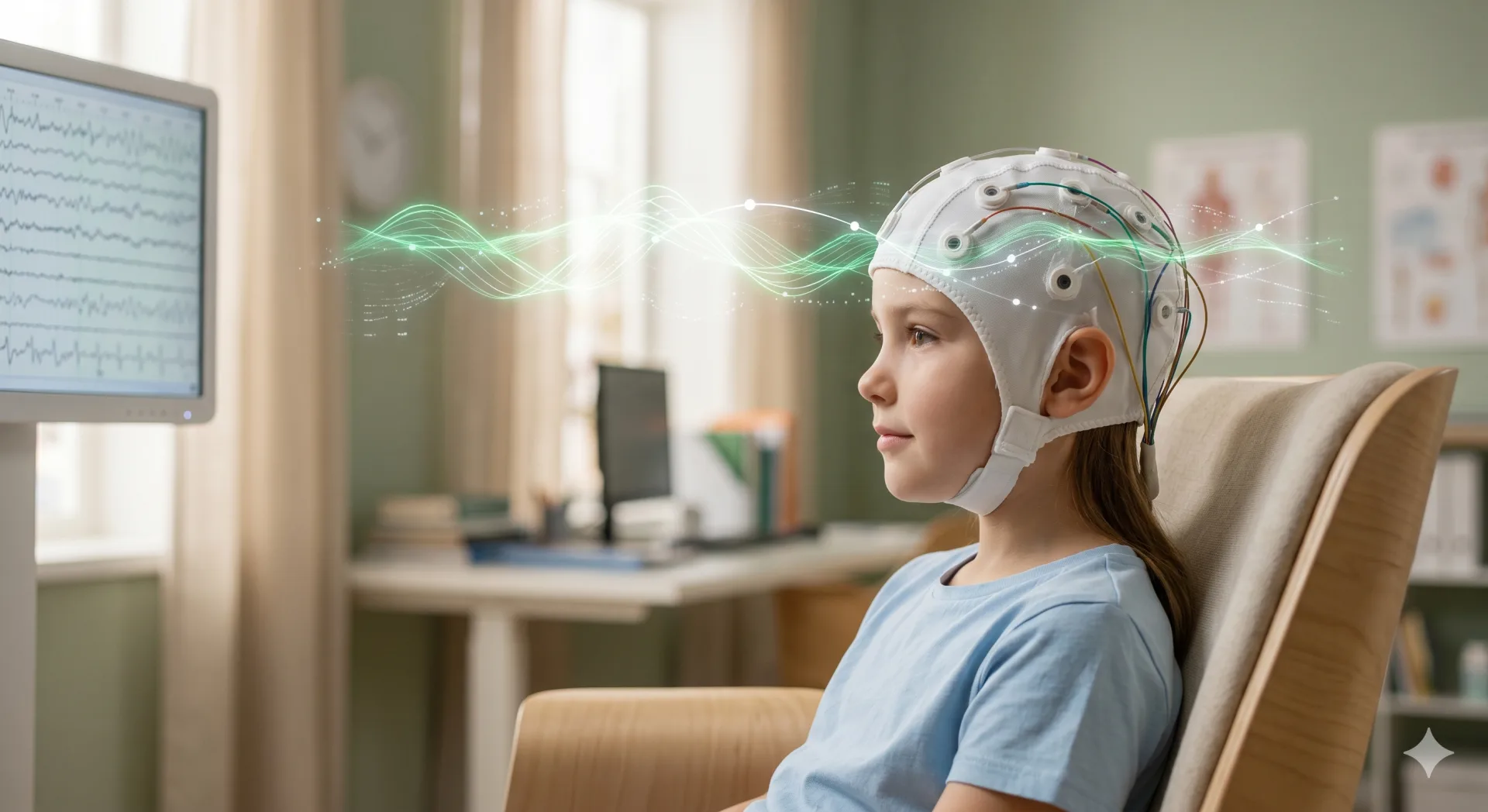
What Is QEEG?
Quantitative EEG (QEEG) is an advanced neuroimaging technique that goes far beyond conventional EEG. While standard EEG displays raw waveforms for a clinician to visually inspect, QEEG processes the same data with sophisticated statistical software to produce a detailed numerical profile of brain activity.
The resulting brain map reveals how different regions communicate with each other, where electrical activity is elevated or suppressed, and how individual patterns compare to large normative databases of healthy individuals. This comparison enables clinically meaningful, evidence-based interpretation.
The procedure is completely painless, free from radiation or contrast agents, and safe for all age groups including young children.
How We Use QEEG
Before initiating stem cell therapy, we establish a detailed neural baseline for each patient. This individualized profile guides the design of the therapy protocol and provides a reference point for measuring change throughout the program.
QEEG is repeated at defined intervals during and following therapy. This objective monitoring allows us to observe whether brainwave patterns are shifting toward healthier norms and to document the neurological effects of therapy with measurable data.
No two brains are alike. By identifying which neural networks are underactive, overactive, or dysconnected, QEEG findings allow us to tailor the therapeutic approach to each individual's unique neurological signature.
What Does QEEG Measure?
QEEG analysis captures six key dimensions of brain electrical activity:
Brainwave Frequency Bands
Delta (0–4 Hz), Theta (4–8 Hz), Alpha (8–12 Hz), and Beta (12–30 Hz) power distributions are analyzed and correlated with states of attention, sleep, relaxation, and cognitive load.
Cortical Connectivity
The degree to which different brain regions work in coordination is assessed. Reduced long-range connectivity is commonly seen in social and communication difficulties, while excess local connectivity may underlie repetitive behaviors.
Hemispheric Asymmetry
Differences in electrical power between the left and right hemispheres are quantified. Abnormal asymmetry patterns have been linked to language delays and emotional regulation difficulties.
Peak Frequency
The dominant frequency in each brain region is identified. Alpha peak frequency in particular serves as an important biomarker of cognitive processing speed and attentional capacity.
Coherence
Coherence measures how synchronously two brain regions oscillate at a given frequency. Both reduced and excessive coherence patterns have been documented in autism spectrum disorder across multiple research studies.
Amplitude (Power Density)
The strength of the electrical signal at each frequency is measured in absolute and relative terms. Abnormal amplitude profiles can serve as objective markers of neurodevelopmental conditions, attention deficits, and anxiety.
The QEEG Process
Preparation
Our team provides clear instructions at least 24 hours in advance, including hair washing and guidance on any relevant medications or caffeine. For children, a child-friendly environment is prepared and families are fully guided through the process.
Electrode Placement
Between 19 and 32 electrodes are placed on the scalp using the standard 10-20 international system and conductive gel. No electricity is sent into the brain — the electrodes only passively record existing neural activity.
Recording (20 Minutes)
The patient rests quietly while brainwave activity is recorded for approximately 20 minutes. Data is collected under multiple conditions, including eyes-open and eyes-closed states, to capture a complete picture of brain function.
Analysis and Report
Raw data is processed and compared to a normative database, producing color-coded brain maps and statistical reports. Our neurology specialist reviews the findings and translates them into clinically meaningful language for the family.
QEEG and Autism Spectrum Disorder
In autism spectrum disorder (ASD), neural network connectivity and brainwave activity patterns differ meaningfully from those seen in neurotypical development. QEEG makes these differences visible, quantifiable, and clinically actionable.
Commonly observed QEEG findings in ASD include elevated frontal theta activity (associated with attention difficulties), reduced long-range connectivity (associated with social-communication challenges), abnormal alpha asymmetry, and right frontal hypoactivity. Because these profiles vary substantially between individuals, QEEG is essential for tailoring therapy rather than applying a one-size-fits-all approach.
Following stem cell therapy, serial QEEG assessments have revealed improvements in connectivity metrics, reductions in excess theta power, and more normalized coherence patterns in a number of our patients. These objective findings give families and clinicians a tangible, data-driven lens through which to evaluate therapeutic progress.
Frequently Asked Questions
Add an Objective Scientific Dimension to Diagnosis and Therapy
QEEG brain mapping is an integrated component of our autism program. Speak with our specialists to begin a personalized, evidence-based journey for your child.
Explore the Autism Program